General Eye Check Up
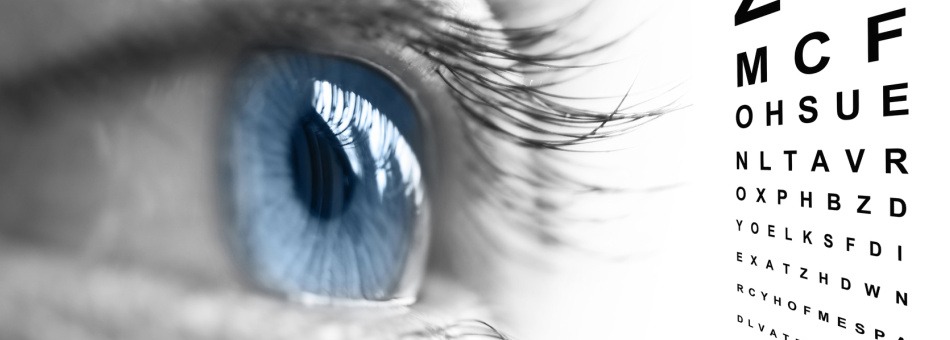
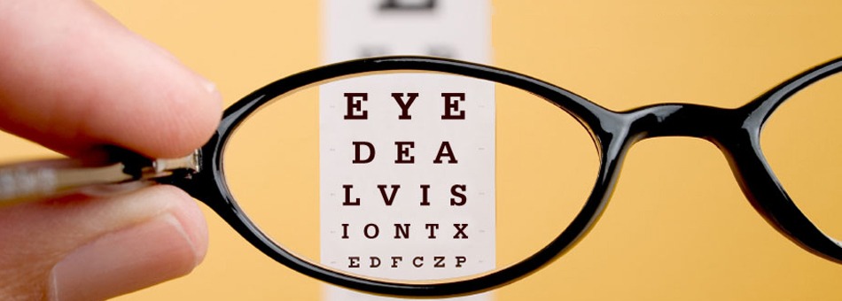
A basic eye exam usually begins with taking a medical history and asking questions about the patient’s general health and past and current eye problems. During the examination, three areas of eye function are assessed: visual acuity and refraction, binocular vision (how the eyes work together), and eye health. Every eye exam uses the Snellen chart to test visual acuity.
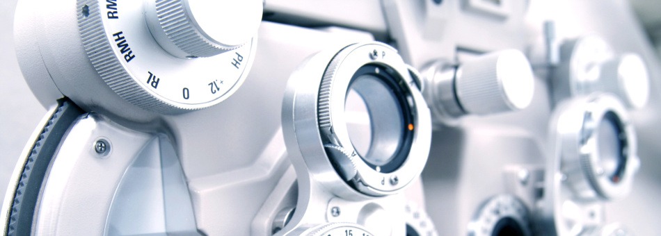
The chart is the one with the big “E.” But that test only identifies 20-30% of vision problems, according to the American Foundation for Vision Awareness. A refraction test helps determine the extent of a patient’s refractive error (nearsightedness, farsightedness, astigmatism), as well as the proper prescription for glasses and contact lenses. A small instrument called a Retinoscope is used to examine light refraction of the eye. The binocular vision assessment is performed to determine how well the two eyes work together. Coordinated eye movement is necessary for accurate depth perception.
During the health assessment, the physician examines the retina the blood vessels of the retina, and the optic nerve head using an ophthalmoscope, a small handheld instrument consisting of a light source and a series of lenses. This examination can help detect signs of many types of diseases. To view the external eye and the more anterior inner eye structures, such as the eyelids, cornea, iris, and lens, a slit lamp microscope is used. For the most thorough look at the internal structures of the eye, the pupils are dilated. Special drops are instilled into the eye that enlarge the pupil and prevent it from constricting when light is shone into the eye during the examination.